abnormal thyroid cancer ultrasound colors
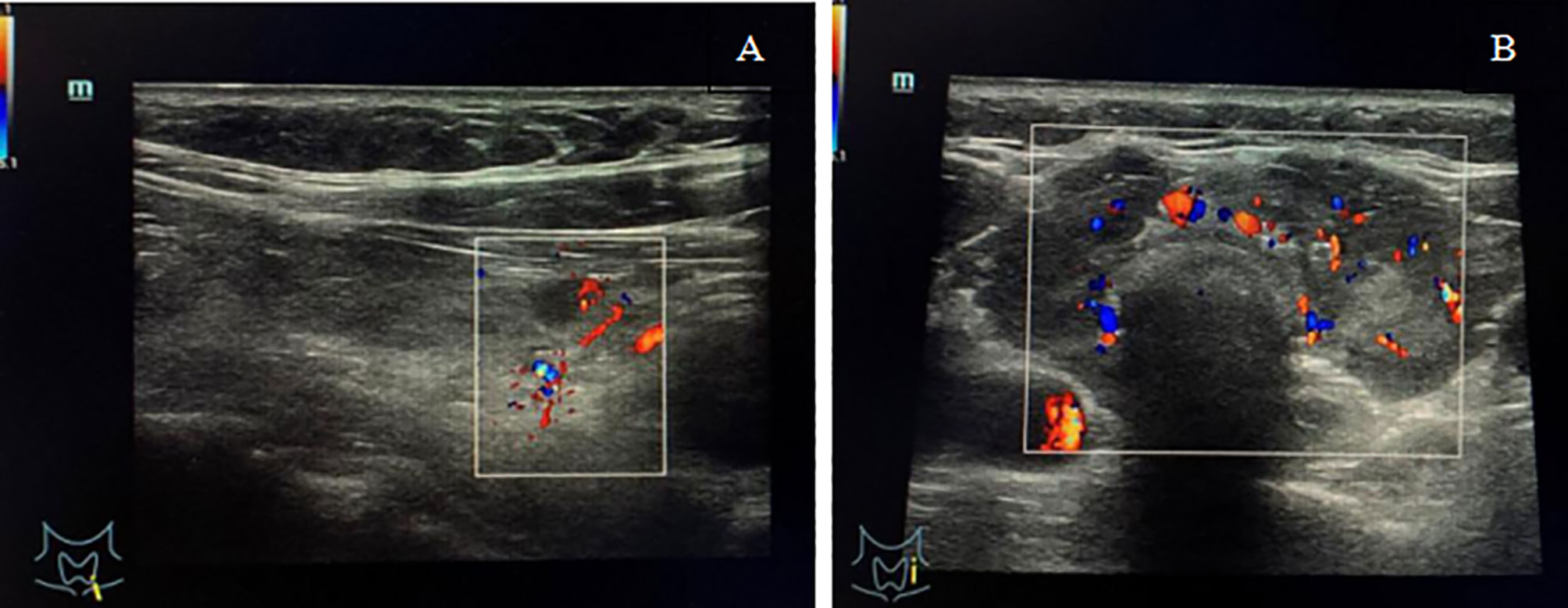
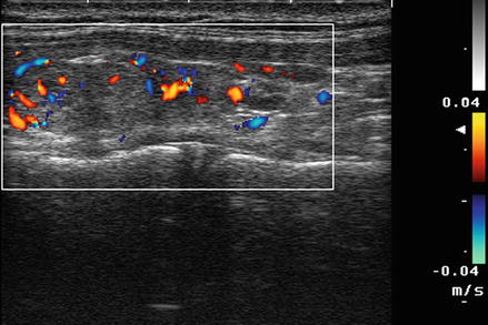
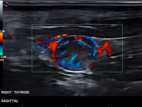
Color on your thyroid ultrasound means that color doppler was applied and blood flow was detected. Vessels in which blood is flowing are colored red for flow in one direction and blue for flow in the other with a color scale that reflects the speed of the flowbecause different.
Thyroid Ultrasound
Thyroid disorder Grayscale ultrasound Color doppler Key.
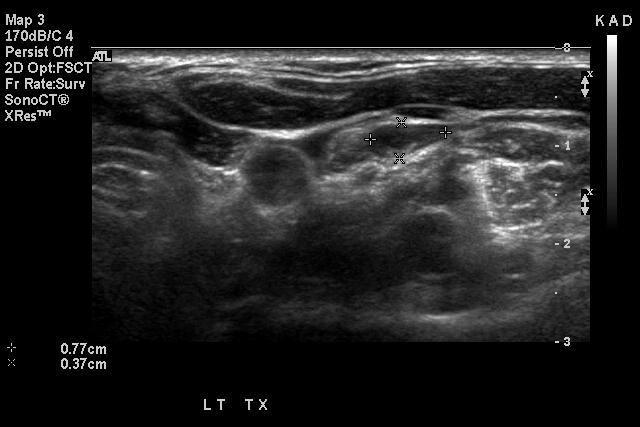
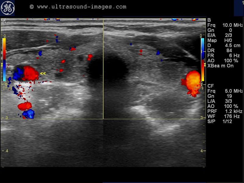
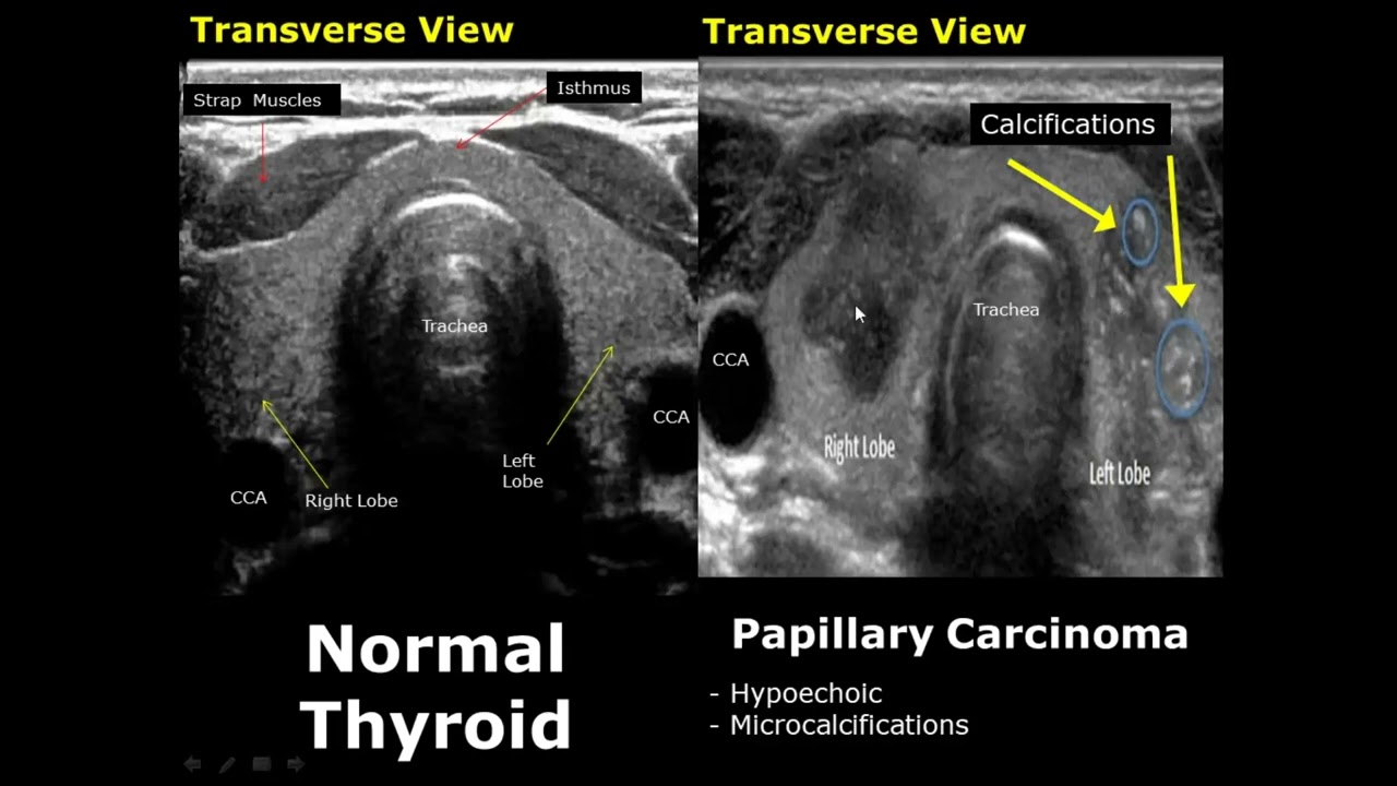
. This nodule shown in red comprises about 80 of the thyroid tissue shown in yellow in this particular area of the thyroid. Thyroid nodular incidentaloma and diffuse thyroid sonographic heterogeneities without discrete nodule. Thyroid nodule thyroid cancer ultrasound colors wednesday august 3 2022 edit.
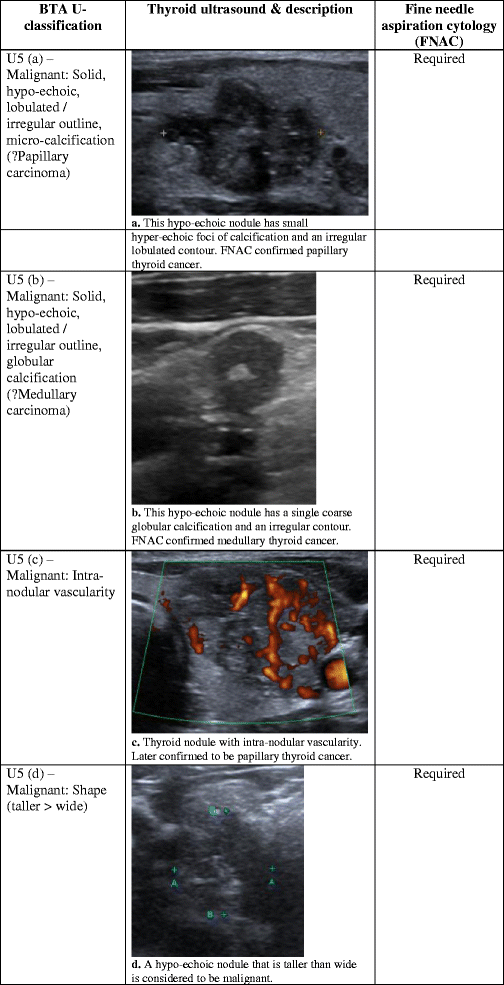
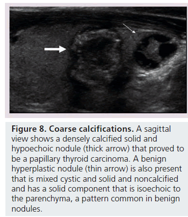
What Abnormal Results Mean Abnormal results may be due to. Pin on tirads us thyroid pin on shitovidka pin on medical imaging pin on endocrinology pin on thyroide pin on sono ultrasound a gallery of high resolution ultrasound. In contrast other studies have shown that ultrasound features such as coarse calcifications more tall than wide irregular borders and increased blood flow within the nodule can be helpful.
In our study we divided incidental thyroid abnormalities in to two groups. Color doppler us uses a computer to convert the doppler. Cysts nodules filled with fluid Enlargement of.
Thyroid Ultrasound Normal Vs Abnormal Image Appearances Comparison Thyroid Pathologies USGTimestampsIntro. Some medical conditions involving the thyroid may have a very similar appearance on a thyroid ultrasound such as Hashimotos thyroiditis and Graves disease. Thyroid hypoechogenicity at ultrasound is a characteristic of autoimmune thyroid diseases with an overlap of this echographic pattern in patients affected by Graves disease or Hashimotos.
It accounts for the majority 70 of all thyroid neoplasms and 85 of all thyroid cancers 24. Thyroid disorder Grayscale ultrasound Color. Cystic appearance hyperechoic punctations calcifications.
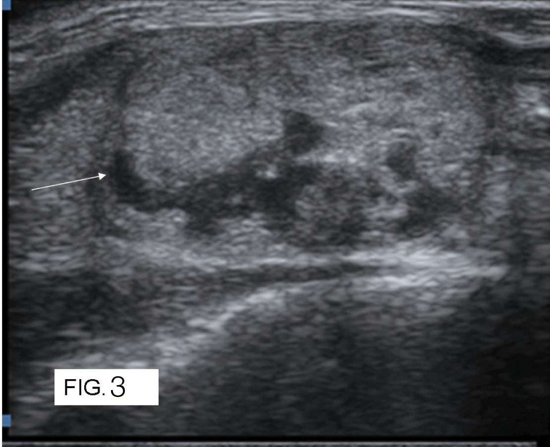
Thyroid Nodule Thyroid Cancer Ultrasound Colors. Features suggestive of metastatic thyroid cancer. A normal result will show that the thyroid has a normal size shape and position.
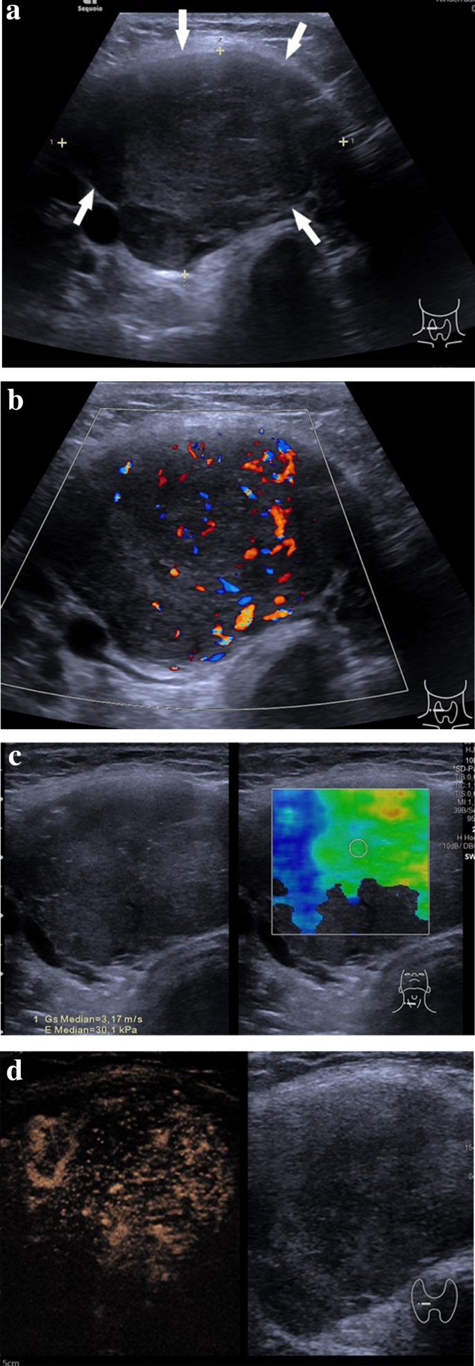
Thyroid ultrasound reveals slight increase in size of lobes from 3 5x12x9 mm to 41x21x 12mm and isthmus is 1. We aimed to investigate the diagnostic value of. The probe is placed on the skin which is at the very top of the picture and sound waves are directed deep into the neck and thyroid.
Ad Find Information For HCPs. Tsh slightly high inspite of synthroidhave hashimotos is the.
Ultrasonography Of Thyroid Nodules A Pictorial Review Insights Into Imaging Full Text

Thyroid Nodule Ultrasound What Is It What Does It Tell Me

Ultrasound Of The Neck There S More To See Than Thyroid Nodules Youtube
Ultrasound Findings Of The Thyroid Gland In Children And Adolescents Springerlink
Thyroid Nodule Sonography Assessment For Risk Of Malignancy
Doppler Ultrasound Radiology Key

Pin On Thyroid
August S Case Of The Month Mivurva

A 52 Year Old Woman With A History Of Hypothyroidism Left Thyroid
A Gallery Of High Resolution Ultrasound Color Doppler 3d Images Thyroid 2

Ultrasonography

Risk For Malignancy Of Thyroid Nodules Comparative Study Between Tirads And Us Based Classification System Sciencedirect
Jcdr Doppler Indices Intranodular Vascularity Ultrasound

Sonography Of Diffuse Thyroid Disease Yuen 2016 Australasian Journal Of Ultrasound In Medicine Wiley Online Library

Thyroid Scintiscanning An Overview Sciencedirect Topics
Frontiers Spontaneous Thyroid Hemorrhage Caused By Langerhans Cell Histiocytosis A Case Report And Literature Review

A 52 Year Old Woman With A History Of Hypothyroidism Left Thyroid
Thyroid Ultrasound Normal Vs Abnormal Image Appearances Comparison Thyroid Pathologies Usg Youtube

Thyroid Gland And Ultrasound Applications Pocket Dentistry